|
|
|
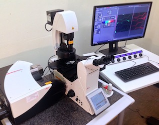
LEICA TCS-SP8 CONFOCAL
LSM
|
INSTRUMENTATION
Confocal Laser Scanning Microscope with:
- Leica DMi8 Advanced inverted light microscope with motorized stage
- LED bright field and EL6000 fluorescence light sources
- 4 laser excitation wavelengths (nm): 405, 488, 552, and 638
- 2 PMT SP fluorescence detectors, one HyD SP detector, transmitted light detector
- LAS-X Software
|
|
|
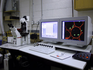
LEICA TCS-SP2 CONFOCAL
LSM
|
 |
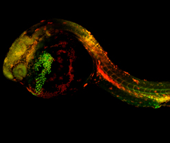
Zebrafish image: Dr. Bryan Crawford, Biology, UNB |
INSTRUMENTATION
Confocal Laser Scanning Microscope with:
- Leica DM IRE2 inverted stand IRE2 light microscope.
- Wide selection of objective lenses including
multi-immersion and long working-distance.
- 4 lasers; Argon/Krypton; HeNe (green); HeNe (orange);
HeNe (red).
- 8 excitation wavelengths: 457, 476, 488, 496,
514, 543, 594, 633.
- 5 detectors - 4 fluorescent channels plus one
transmitted light detector.
- AOBS (programmable beam splitter).
- Physiology and materials software.
|
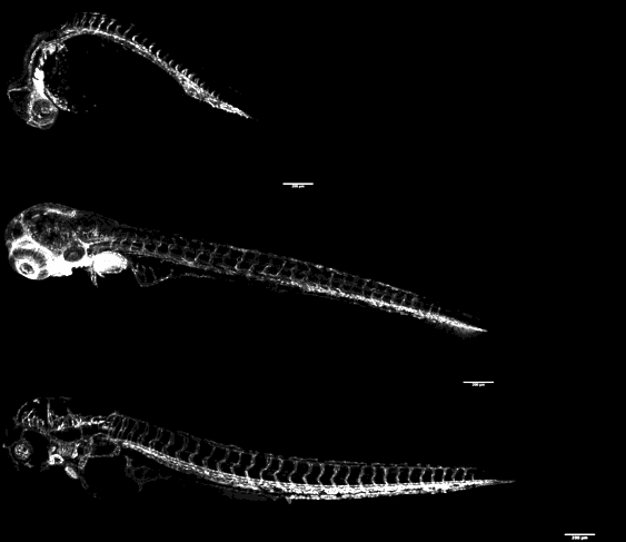 Rotating projections of composite confocal stacks
through transgenic zebrafish embryos.
Rotating projections of composite confocal stacks
through transgenic zebrafish embryos.
Green
fluorescent protein (GFP) in developing endothelial
cells.
Top embryo, 24 hpf; middle embryo 48 hpf; bottom
embryo, 96 hpf.
Class of Biology 3181, UNB Biology, Winter 2011.
|
|
|
|
|
|