|
|
|
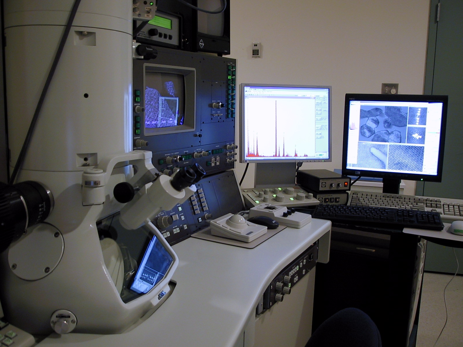
JEOL 2011 STEM |

|

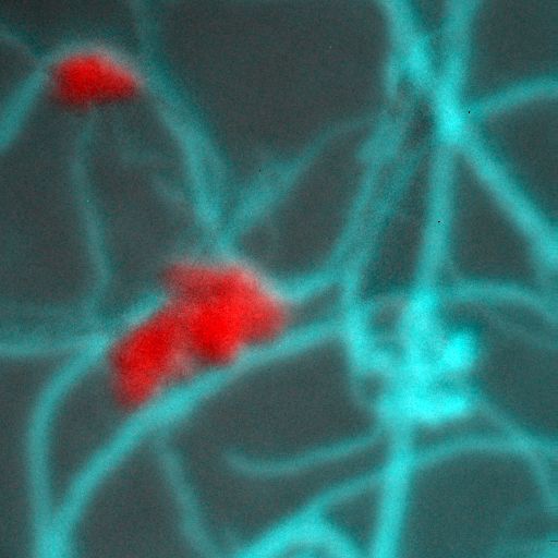
EELS Map: Louise Weaver, Ph.D., Microscopy
& Microanalysis Facility, UNB |
(SCANNING) TRANSMISSION ELECTRON MICROSCOPY
The (S)TEM can be used to examine
internal structure and composition of thin, thinned,
or sectioned specimens. Convergent beam electron
diffraction provides information on crystal structure
and crystallography. STEM and high-angle annular
dark field gives information on microstructure,
atomic structure and atomic number. Energy dispersive
x-ray spectroscopy permits submicrometer elemental
identification and compositional analysis. The addition
of electron energy loss spectroscopy and energy-filtered
imaging allows for the detection of elements at
higher spatial resolution, phase identification
and bonding information. In summary, the (S)TEM
with X-ray and EELS spectroscopy permits further
characterization of both organic and inorganic materials.
IMAGING TECHNIQUES
- Brightfield – Conventional mode (CTEM),
STEM mode
- Darkfield - High Angle Annular Darkfield (HAADF)
- Electron Diffraction – High Resolution
(HR), Selected Area (SA), High Dispersion(HD)
- Energy filtered TEM
ANALYTICAL MODES
- Elemental recognition and phase identification
- Quantitative compositional analysis
- Digital x-ray maps and linescans
- Atomic structure - Z-contrast imaging (HAADF)
- Energy-filtered TEM (EFTEM)
- Bonding analysis - electron energy loss spectroscopy
(EELS)
INSTRUMENTATION
JEOL 2011 SCANNING TRANSMISSION ELECTRON MICROSCOPE
with:
- EDAX (Genesis) Energy Dispersive X-ray system
- Gatan Imaging Filter (GIF 2000) with EFTEM and
EELS capabilities
- Gatan 4k x 4k Ultrascan digital camera
- Gatan DualVision digital camera
- Gatan Cryo-transfer and cooling holder
- JEOL double-tilt analytical holder
|
|
|
|